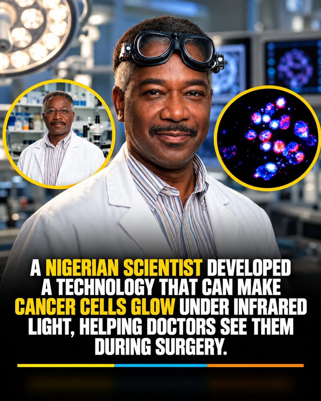
The Nigerian-Born Scientist Helping Surgeons See Cancer Cells Glow 🔬✨
The Nigerian-Born Scientist Helping Surgeons See Cancer Cells Glow 🔬✨
It sounds like something from a science-fiction movie: cancerous tissue glowing during surgery while healthy tissue remains comparatively dark. But this idea is rooted in real medical imaging research.
At the center of the technology is Samuel Achilefu, a Nigerian-born biomedical scientist whose work has focused on using near-infrared molecular imaging to help surgeons visualize cancer during operations. The Optical Society recognizes Achilefu for pioneering work in near-infrared molecular imaging and image-guided cancer surgery.
How can cancer cells “glow”?
The technology doesn’t mean cancer cells naturally produce visible light.
Instead, a specially designed fluorescent contrast agent can be administered to a patient. The agent is designed to interact with or accumulate in targeted cancerous tissue. When illuminated with particular wavelengths of light, the targeted tissue produces a signal that specialized imaging equipment can detect.
The signal can then be displayed to the surgeon, helping distinguish areas of concern from surrounding tissue. Recent reports describing Achilefu’s work say his medical-vision goggles combine fluorescence imaging, near-infrared light and specialized cameras to provide surgeons with enhanced visual information during an operation.
Why could this matter?
One of the challenges in cancer surgery is determining exactly where a tumor ends and healthy tissue begins. Surgeons already use imaging, pathology, visual inspection and their clinical judgment, but microscopic or difficult-to-see tumor tissue can sometimes be challenging to identify.
Image-guided surgery is intended to provide additional information in real time, potentially helping surgeons locate targeted cancerous tissue and identify areas that might otherwise be difficult to distinguish.
Importantly, this technology should not be described as a universal cure for cancer or as a system that automatically detects every cancer cell. Its role is to assist medical teams with visualization, and its usefulness depends on the particular cancer, imaging agent, technology and clinical setting.
From Nigeria to global medical research
Achilefu was born in Nigeria and went on to build an international career in biomedical engineering, radiology and cancer imaging. His work has contributed to the broader field of molecular imaging and image-guided cancer treatment.
The idea behind his research is remarkably simple to understand: if surgeons can see cancer more clearly, they may have better information while removing it.
What once sounded like science fiction—using light and specialized imaging technology to make cancerous tissue stand out—is now part of a real and rapidly developing area of medical research.
And while more research and clinical validation are needed for different applications, the technology represents an intriguing example of how optics, chemistry, engineering and medicine can come together to tackle one of surgery’s most difficult challenges. 🔬✨